


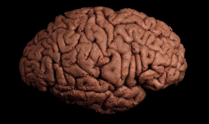
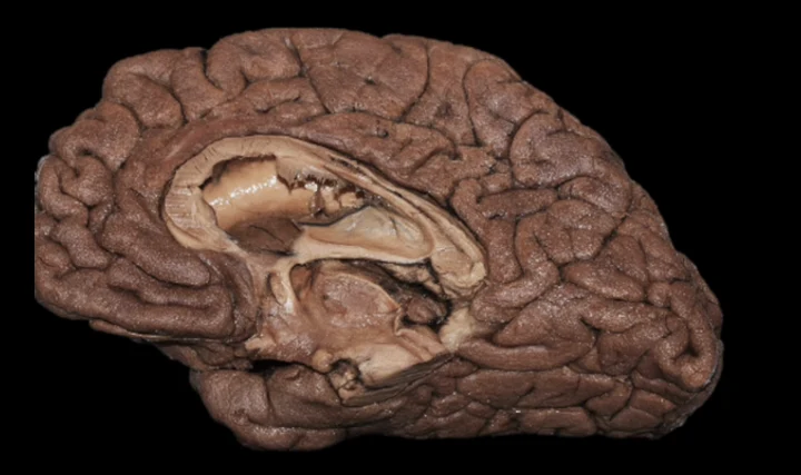
Virtual White matter dissection
Virtual dissection offers a valuable tool for students, residents, neurosurgeons, and neuroscientists interested in studying white matter dissection. Photogrammetric models can be explored in 3D in a stratigraphic sequence, faithfully reproducing, within a virtual reality environment, what can normally be observed in the anatomical laboratory. The slider allows users to select the dissection plane of interest, presented in the sequence of a classical dissection. Individual structures can be explored using the labels and definitions available in the adjacent drop-down menu.
White Matter fibers Dissection: a brief history
The history of the developments and discoveries that have shaped our current understanding of the macroscopic anatomy of the cerebral white matter is a rich one. Several distinguished philosophers and scientists have contributed to this field, including Galen, Piccolomini, Malpighi, Varolio, Willis, Steno, Vieussens, Vicq d’Azyr, Reil, Gall, Burdach, Meynert, Mayo, Arnold, Foville and Gratiolet, to name just a few.
In the nineteenth century, however, there was a radical shift in focus. Most scientists turned their attention from the macroscopic to the microscopic anatomy of the human cerebral white matter, so that white matter dissection was used mainly for teaching purposes throughout the twentieth century. In the 1930s, Mr Klingler, an anatomical demonstrator in Professor Ludwig’s anatomical laboratory in Basel, developed a particular interest and talent for white matter dissection.
His aim was to provide students at the University of Basel with three-dimensional models that would “free students from the task of mentally reconstructing structures after examining numerous sections, a task that all too often proves unsuccessful.” He also made a slight modification to the dissection protocol by routinely freezing brain specimens, a detail he had adopted during his time in Vienna at Professor Pernkopf’s anatomical laboratory.
Mr Klingler’s talent resulted in a remarkable atlas of white matter fibre dissection: the Atlas cerebri humani, which remains unsurpassed in its level of detail and the perfection of its execution. Several students benefited from Mr Klingler’s expertise, including the young Professor Yaşargil, who visited Professor Ludwig’s laboratory for three months in the 1940s. Thereafter, he regularly sought out Mr Klingler to show him his improvements in dissection techniques, usually receiving the reply: “You are getting better and better…”
In the second half of the twentieth century, the technique fell into obscurity, and only a few studies on the subject appeared in the medical literature. In the 1990s, one of the young neurosurgeons visiting Professor Yaşargil in Zurich was struck by the beauty and precision of the dissected specimens that Professor Yaşargil kept in his office. At Professor Yaşargil’s suggestion, he subsequently visited the Anatomical Museum in Basel to admire Mr Klingler’s original specimens.
This young colleague, now Professor Uğur Türe, published a paper in 2000 describing, step by step, how to perform the lateral dissection of the brain. This work marked a milestone in the history of white matter dissection and generated considerable interest in the subject within the scientific community.
Credits
The VIRTUAL ANATOMY PLATFORM was conceived and developed by Marcello Magnani, MD, under the patronage of the MICN Lab.
We gratefully acknowledge the Institute of Anatomy, University of Zurich (Campus Irchel), and in particular Giovanni Colacicco and Beatrice Astrid Lauber, for their cooperation.
References
- Türe U, Yaşargil DC, Al-Mefty O, Yaşargil MG. Topographic anatomy of the insular region. J Neurosurg. 1999;90(4):720–733. doi:10.3171/jns.1999.90.4.0720
- Türe U, Yaşargil MG, Friedman AH, Al-Mefty O. Fiber dissection technique: lateral aspect of the brain. Neurosurgery. 2000;47(2):417–427.
- Yaşargil MG, Türe U, Yaşargil DC. Surgical anatomy of supratentorial midline lesions. Neurosurg Focus. 2005;18(6B):E1. doi:10.3171/foc.2005.18.6.14
- Serra C, Türe U, Krayenbühl N, Şengül G, Yaşargil DC, Yaşargil MG. Topographic classification of the thalamus surfaces related to microneurosurgery: a white matter fiber microdissection study. World Neurosurg. 2017;97:438–452. doi:10.1016/j.wneu.2016.09.101
- Goga C, Serra C, Türe U. Microsurgical anatomy and the inner architecture of the retrocommissural portion of the hippocampal formation demonstrated through fiber microdissection. J Neurosurg. 2025;142(4):1085–1098. doi:10.3171/2024.6.JNS232987
- Zanuttini L, Staartjes VE, Chen S-C, Lin C-F, Hsu SPC, Martins C, Wen HT, Serra C, Türe U. White matter microdissection of the lateral aspect of the brain: 2-dimensional video demonstration. Acta Neurochir. 2025;167:215. doi:10.1007/s00701-025-06635-z
- Piazza A, Stumpo V, Staartjes V, Colacicco G, de Notaris M, Frati A, Agosti E, Regli L, Krayenbühl N, Serra C, Türe U. Virtual human white matter dissection: a stratigraphic layer-by-layer dissection of human brain in photogrammetry. Oper Neurosurg. 2026. doi:10.1227/ons.0000000000001946
- Zanuttini L, Staartjes VE, Menna G, Chen S-C, Lin C-F, Hsu SPC, Martins C, Wen HT, Colacicco G, Kadri PAS, Krayenbühl N, Serra C, Türe U. White matter microdissection of the medial aspect of the brain: 2-dimensional video demonstration. Acta Neurochir. 2026;168:55. doi:10.1007/s00701-026-06791-w